

Volume 10, Issue 3 (11-2024)
J Sport Biomech 2024, 10(3): 230-240 |
Back to browse issues page


![]()
![]()
![]()
Download citation:
BibTeX | RIS | EndNote | Medlars | ProCite | Reference Manager | RefWorks
Send citation to:


BibTeX | RIS | EndNote | Medlars | ProCite | Reference Manager | RefWorks
Send citation to:
Shahbazi M A, Jalalvand A. Comparison of Spatiotemporal Gait Variables Between Healthy Individuals and Patients with Heel Spur During Walking. J Sport Biomech 2024; 10 (3) :230-240
URL: http://biomechanics.iauh.ac.ir/article-1-352-en.html
URL: http://biomechanics.iauh.ac.ir/article-1-352-en.html


1- Department of Sport Biomechanics, Hamedan Branch, Islamic Azad University, Hamedan, Iran.
Full-Text [PDF 1629 kb]
(603 Downloads)
| Abstract (HTML) (2468 Views)
Full-Text: (1008 Views)
Extended Abstract
1. Introduction
A heel spur is a small, pointed bony protrusion that forms beneath the heel, near the attachment site of the ligaments and the plantar fascia to the heel bone (calcaneus). This condition typically arises from excessive pressure, inflammation, or repetitive stretching of the plantar fascia (1). Walking on uneven surfaces places significant stress on the sole of the foot, exacerbating the problems associated with heel spurs. Heel spurs can alter natural posture during walking and, over time, may lead to complications such as back pain and knee pain (5). These issues are frequently observed in professional athletes, individuals with obesity, and those who wear inappropriate footwear. In most cases, there is no history of trauma, and the pain is most pronounced upon rising from sleep. While the discomfort tends to diminish after taking a few steps, it intensifies with prolonged activity, particularly after completing daily tasks. Clinical examination often reveals tenderness in the anterior-medial region of the inferior heel surface (7). Intense physical activity worsens the pain associated with heel spurs and impacts kinematic parameters, including knee flexion angles during load-bearing, ankle dorsiflexion during load-bearing, and ankle plantarflexion (4). However, the effect of this condition on spatiotemporal gait parameters remains underexplored. Given that musculoskeletal disorders of the foot can impair lower limb function (8) and induce various changes in biomechanical parameters (9), analyzing the kinematic changes, particularly spatiotemporal parameters, in such disorders is essential for designing therapeutic interventions aimed at improving recovery and enhancing functionality.
2. Methods
The study population consisted of patients with heel spurs and their healthy counterparts. A total of 30 participants were selected through convenience sampling and divided into two groups of 15 based on statistical calculations performed using G*Power software. The inclusion criteria required male participants aged 30–49 years with a diagnosis of heel spurs and a minimum pain score of 3 on the numeric-visual scale. Exclusion criteria included the presence of bone or neurological disorders, diabetes, or skeletal deformities. Spatiotemporal gait parameters were measured during walking at a sampling frequency of 100 Hz using Vicon high-speed cameras. The data were assessed for normality using the Shapiro-Wilk test, and statistical analysis was conducted using an independent t-test in SPSS software.
3. Results
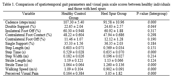
Individuals with plantar fasciitis demonstrated a higher percentage of double support, step time, stride time, and visual pain intensity, along with lower cadence and walking speed compared to the healthy control group (Table 1).
4. Conclusion
Individuals with heel spurs exhibit lower step cadence compared to the healthy group. One primary reason for this reduction is the avoidance of pain. Patients with heel spurs often experience heel pain during walking and other activities (11). To alleviate discomfort, they tend to walk more slowly, take shorter steps, and spend less time on the affected foot. These pathomechanical adaptations naturally result in reduced step cadence as patients adopt these strategies to protect the painful area (11, 12). Individuals with heel spurs also demonstrate a higher double support time compared to the healthy group. Heel pain during weight-bearing activities, especially walking, leads patients to spend less time on the affected foot and distribute their weight more evenly between both feet. Consequently, the duration of the double support phase, when both feet are in contact with the ground, increases (12). Step time is longer in individuals with heel spurs compared to the healthy group. Patients may develop an antalgic gait, a walking pattern that minimizes pain. This pattern often involves spending less time on the affected foot, leading to asymmetry in step time and a decrease in overall walking speed (12). Stride time is also longer in individuals with heel spurs. Pain in the heel area can prompt individuals to adjust how their foot contacts the ground. Instead of landing on the heel, which may exacerbate plantar fascia strain, they modify their landing method, slowing down the gait cycle (15). Walking speed is lower in individuals with heel spurs compared to the healthy group. Due to weaker postural control during walking, these individuals reduce their walking speed to maintain balance (11). Lastly, individuals with heel spurs report higher levels of visual pain intensity compared to the healthy control group. While the heel spur itself may not always be the primary source of pain, its presence is often associated with inflammation and irritation of the surrounding soft tissues, particularly the plantar fascia. This inflammation can result in significant discomfort (17, 18).
Ethical Considerations
Compliance with ethical guidelines
There were no ethical considerations to be addressed in this research.
Funding
This research did not receive any financial support from government, private, or non-profit organizations.
Authors' contributions
All authors contributed equally to preparing the article.
Conflicts of interest
The authors declare that there are no conflicts of interest associated with this article.
A heel spur is a small, pointed bony protrusion that forms beneath the heel, near the attachment site of the ligaments and the plantar fascia to the heel bone (calcaneus). This condition typically arises from excessive pressure, inflammation, or repetitive stretching of the plantar fascia (1). Walking on uneven surfaces places significant stress on the sole of the foot, exacerbating the problems associated with heel spurs. Heel spurs can alter natural posture during walking and, over time, may lead to complications such as back pain and knee pain (5). These issues are frequently observed in professional athletes, individuals with obesity, and those who wear inappropriate footwear. In most cases, there is no history of trauma, and the pain is most pronounced upon rising from sleep. While the discomfort tends to diminish after taking a few steps, it intensifies with prolonged activity, particularly after completing daily tasks. Clinical examination often reveals tenderness in the anterior-medial region of the inferior heel surface (7). Intense physical activity worsens the pain associated with heel spurs and impacts kinematic parameters, including knee flexion angles during load-bearing, ankle dorsiflexion during load-bearing, and ankle plantarflexion (4). However, the effect of this condition on spatiotemporal gait parameters remains underexplored. Given that musculoskeletal disorders of the foot can impair lower limb function (8) and induce various changes in biomechanical parameters (9), analyzing the kinematic changes, particularly spatiotemporal parameters, in such disorders is essential for designing therapeutic interventions aimed at improving recovery and enhancing functionality.
2. Methods
The study population consisted of patients with heel spurs and their healthy counterparts. A total of 30 participants were selected through convenience sampling and divided into two groups of 15 based on statistical calculations performed using G*Power software. The inclusion criteria required male participants aged 30–49 years with a diagnosis of heel spurs and a minimum pain score of 3 on the numeric-visual scale. Exclusion criteria included the presence of bone or neurological disorders, diabetes, or skeletal deformities. Spatiotemporal gait parameters were measured during walking at a sampling frequency of 100 Hz using Vicon high-speed cameras. The data were assessed for normality using the Shapiro-Wilk test, and statistical analysis was conducted using an independent t-test in SPSS software.
3. Results
Individuals with plantar fasciitis demonstrated a higher percentage of double support, step time, stride time, and visual pain intensity, along with lower cadence and walking speed compared to the healthy control group (Table 1).
4. Conclusion
Individuals with heel spurs exhibit lower step cadence compared to the healthy group. One primary reason for this reduction is the avoidance of pain. Patients with heel spurs often experience heel pain during walking and other activities (11). To alleviate discomfort, they tend to walk more slowly, take shorter steps, and spend less time on the affected foot. These pathomechanical adaptations naturally result in reduced step cadence as patients adopt these strategies to protect the painful area (11, 12). Individuals with heel spurs also demonstrate a higher double support time compared to the healthy group. Heel pain during weight-bearing activities, especially walking, leads patients to spend less time on the affected foot and distribute their weight more evenly between both feet. Consequently, the duration of the double support phase, when both feet are in contact with the ground, increases (12). Step time is longer in individuals with heel spurs compared to the healthy group. Patients may develop an antalgic gait, a walking pattern that minimizes pain. This pattern often involves spending less time on the affected foot, leading to asymmetry in step time and a decrease in overall walking speed (12). Stride time is also longer in individuals with heel spurs. Pain in the heel area can prompt individuals to adjust how their foot contacts the ground. Instead of landing on the heel, which may exacerbate plantar fascia strain, they modify their landing method, slowing down the gait cycle (15). Walking speed is lower in individuals with heel spurs compared to the healthy group. Due to weaker postural control during walking, these individuals reduce their walking speed to maintain balance (11). Lastly, individuals with heel spurs report higher levels of visual pain intensity compared to the healthy control group. While the heel spur itself may not always be the primary source of pain, its presence is often associated with inflammation and irritation of the surrounding soft tissues, particularly the plantar fascia. This inflammation can result in significant discomfort (17, 18).
Ethical Considerations
Compliance with ethical guidelines
There were no ethical considerations to be addressed in this research.
Funding
This research did not receive any financial support from government, private, or non-profit organizations.
Authors' contributions
All authors contributed equally to preparing the article.
Conflicts of interest
The authors declare that there are no conflicts of interest associated with this article.
Type of Study: Research |
Subject:
Special
Received: 2024/12/14 | Accepted: 2024/12/28 | Published: 2024/12/28
Received: 2024/12/14 | Accepted: 2024/12/28 | Published: 2024/12/28
References
1. Azar FM, Canale ST, Beaty JH. Campbell's Operative Orthopaedics, E-Book: Elsevier Health Sciences; 2020.
2. Haake M, Buch M, Schoellner C, Goebel F, Vogel M, Mueller I, et al. Extracorporeal shock wave therapy for plantar fasciitis: randomised controlled multicentre trial. Bmj. 2003;327(7406):75. [DOI:10.1136/bmj.327.7406.75] [PMID]
3. Riddle DL, Pulisic M, Pidcoe P, Johnson RE. Risk factors for plantar fasciitis: a matched case-control study. JBJS. 2003;85(5):872-7. [DOI:10.2106/00004623-200305000-00015] [PMID]
4. Wearing SC, Smeathers JE, Urry SR. The effect of plantar fasciitis on vertical foot-ground reaction force. Clinical Orthopaedics and Related Research (1976-2007). 2003;409:175-85. [DOI:10.1097/01.blo.0000057989.41099.d8] [PMID]
5. Zahnreich S, Rösler H-P, Schwanbeck C, Karle H, Schmidberger H. Radiation-induced DNA double-strand breaks in peripheral leukocytes and therapeutic response of heel spur patients treated by orthovoltage X-rays or a linear accelerator. Strahlentherapie und Onkologie. 2020;196:1116-27. [DOI:10.1007/s00066-020-01662-4] [PMID]
6. Altuntas E, Uzun A. The Relationship Between Age-Related Incidences of Heel Spur With Sex and Side. International Journal of Morphology. 2022;40(2):369-375. [DOI:10.4067/S0717-95022022000200369]
7. Nawaz A, Khan U, Zaheer A, Shahzadi A. Prevalence of heel spur in teachers. Pakistan Journal of Physical Therapy (PJPT). 2022. [DOI:10.52229/pjpt.v5i1.1667]
8. Tanaka C, Farah EA. Anatomia funcional das cadeias musculares. Fisioterapia e Pesquisa. 1997;4(1):39-40.
9. Williams III DS, McClay IS, Hamill J, Buchanan TS. Lower extremity kinematic and kinetic differences in runners with high and low arches. Journal of applied biomechanics. 2001;17(2):153-63. [DOI:10.1123/jab.17.2.153]
10. Faul F, Erdfelder E, Lang A-G, Buchner A. G* Power 3: A flexible statistical power analysis program for the social, behavioral, and biomedical sciences. Behavior research methods. 2007;39(2):175-91. [DOI:10.3758/BF03193146] [PMID]
11. Richer L, Fortin E, Gagnon G, Ngomo S, Fernandes KBP, Cortis C, et al. Impact of plantar fasciitis on postural control and walking in young middle-aged adults. The Foot. 2022;53:101951. [DOI:10.1016/j.foot.2022.101951] [PMID]
12. Thong-On S, Bovonsunthonchai S, Vachalathiti R, Intiravoranont W, Suwannarat S, Smith R. Effects of strengthening and stretching exercises on the temporospatial gait parameters in patients with plantar fasciitis: A randomized controlled trial. Annals of rehabilitation medicine. 2019;43(6):662-76. [DOI:10.5535/arm.2019.43.6.662] [PMID]
13. Yoo SD, Kim HS, Lee JH, Yun DH, Kim DH, Chon J, et al. Biomechanical parameters in plantar fasciitis measured by gait analysis system with pressure sensor. Annals of rehabilitation medicine. 2017;41(6):979-89. [DOI:10.5535/arm.2017.41.6.979] [PMID]
14. Chandler T, Kibler W. A biomechanical approach to the prevention, treatment and rehabilitation of plantar fasciitis. Sports Medicine. 1993;15:344-52. [DOI:10.2165/00007256-199315050-00006] [PMID]
15. Martin RL, Davenport TE, Reischl SF, McPoil TG, Matheson JW, Wukich DK, et al. Heel pain-plantar fasciitis: revision 2014. Journal of Orthopaedic & Sports Physical Therapy. 2014;44(11):A1-A33. [DOI:10.2519/jospt.2014.0303] [PMID]
16. El-helw MR, Fathallah MM, El Zohiery AK, Mostafa NH. Comparison between the effect of Steroid Injection, Prolotherapy Local Injection and Extracorporal Shock Wave Therapy in Treatment of Symptomatic Calcaneal Spur. QJM: An International Journal of Medicine. 2024;117(Supplement_1):hcae070. 535. [DOI:10.1093/qjmed/hcae070.535]
17. Beyzadeoğlu M, Sağer Ö, Dinçoğlan F, Demiral S, Uysal B, Gamsız H, et al. Evaluation of the radiotherapeutic management of refractory painful heel spur and plantar fasciitis: a single center experience of 45 years. Evaluation. 2020;62(1):8-13. [DOI:10.4274/gulhane.galenos.2019.730]
18. Okçu M, Tuncay F, Koçak FA, Erden Y, Ayhan MY, Kaya SS. Do the presence, size, and shape of plantar calcaneal spurs have any significance in terms of pain and treatment outcomes in patients with plantar fasciitis? Turkish journal of medical sciences. 2023;53(1):413-9. [DOI:10.55730/1300-0144.5598] [PMID]
| Rights and permissions | |
 |
This work is licensed under a Creative Commons Attribution-NonCommercial 4.0 International License. |
Journal of Sport Biomechanics
Department of Sport Biomechanics, Faculty of Humanities, Islamic Azad University, Hamedan Branch, Prof. Mousivand Blvd, Imam Khomeini Blvd, Hamedan, Iran.
Journal Tel: +98 81 34494042
Website: http://biomechanics.iauh.ac.ir
Email: sportbiomechanics@iauh.ac.ir
