

Volume 12, Issue 3 (11-2026)
J Sport Biomech 2026, 12(3): 546-568 |
Back to browse issues page




Download citation:
BibTeX | RIS | EndNote | Medlars | ProCite | Reference Manager | RefWorks
Send citation to:


BibTeX | RIS | EndNote | Medlars | ProCite | Reference Manager | RefWorks
Send citation to:
Ilbeigi S, Motedayyen H, Yousefi M, Hosseini S N, Hoseinzadeh E. Comparison of the Effects of Exercise Therapy and Orthokine Therapy on Pain Intensity, Joint Stiffness, and Quadriceps Muscle Strength in Middle-Aged Women with Knee Osteoarthritis. J Sport Biomech 2026; 12 (3) :546-568
URL: http://biomechanics.iauh.ac.ir/article-1-433-en.html
URL: http://biomechanics.iauh.ac.ir/article-1-433-en.html
Saeed Ilbeigi *1 

 , Haniyeh Motedayyen1 , Mohammad Yousefi1 , Seyyed Naser Hosseini2 , Effat Hoseinzadeh1
, Haniyeh Motedayyen1 , Mohammad Yousefi1 , Seyyed Naser Hosseini2 , Effat Hoseinzadeh1
1- Department of Sport Sciences, Faculty of Sport Sciences, University of Birjand, Birjand, Iran.
2- Physical Medicine and Rehabilitation Specialist, Mashhad, Iran.
2- Physical Medicine and Rehabilitation Specialist, Mashhad, Iran.
Full-Text [PDF 1975 kb]
(7 Downloads)
| Abstract (HTML) (14 Views)
Full-Text: (7 Views)
1. Introduction
Osteoarthritis (OA) is one of the most common joint disorders, with a considerably higher prevalence in the knee than in any other joint of the human body (1). Compared with osteoarthritis affecting other joints, knee osteoarthritis (KOA) is associated with greater disability and more pronounced clinical manifestations, making it a major global health challenge (2). KOA is a leading cause of pain and functional limitation across all segments of the population, particularly among individuals over 50 years of age. Its prevalence has doubled since the mid-twentieth century and is expected to rise at an even faster rate in the future (3). The mechanisms underlying OA development are complex. One hypothesis suggests that cartilage degradation and the subsequent release of cartilage debris into the synovial fluid trigger inflammatory responses. Another hypothesis proposes that microscopic fractures in the subchondral bone lead to sclerosis and hypertrophy, thereby contributing to cartilage wear (4). In both pathways, the surrounding muscles, tendons, and ligaments weaken and undergo physiological changes, which can result in pain, reduced range of motion, and joint swelling (1, 3). Consequently, patients tend to adopt a more sedentary lifestyle, leading to a substantial decline in their quality of life (3).
To date, no definitive cure for KOA has been established (5), and most currently available interventions have been developed to manage the disease by reducing pain, slowing cartilage degeneration, and improving joint function (1, 3, 6). Current management strategies for knee osteoarthritis include pharmacological and non-pharmacological treatments, stem cell therapy, and joint surgery. Based on the available evidence, pharmacological and non-pharmacological approaches, such as exercise therapy, weight reduction, and the use of orthotic devices, are generally recommended as first-line treatments for patients with knee osteoarthritis (6). Among these, exercise therapy is recognized as one of the cornerstones of conservative management and plays an important role in reducing pain, improving joint function, and controlling disease progression by enhancing muscle strength, improving proprioception, and optimizing neuromuscular responses (7). The National Institute for Health and Care Excellence (NICE) identifies exercise therapy as the primary non-pharmacological intervention for the management of knee osteoarthritis. Evidence indicates that physical activity can alleviate symptoms and improve functional outcomes in patients with knee osteoarthritis by modulating inflammatory and metabolic processes within the joint (8). The findings of several systematic reviews and meta-analyses have demonstrated that exercise therapy can significantly improve muscle function, increase joint range of motion, and reduce pain, thereby enhancing physical function and quality of life in patients with knee osteoarthritis (9). Nevertheless, the implementation of exercise therapy in the management of knee osteoarthritis remains limited or is often delivered with suboptimal quality due to factors such as the prolonged duration required for treatment and therapeutic effectiveness, the absence of a standardized and universally accepted treatment protocol for knee osteoarthritis, physicians’ limited adherence to completing the full course of treatment, and patients’ poor compliance with performing progressive exercise because of pain and fear of physical activity (6, 10).
Conversely, some researchers have reported that pharmacological treatments may be effective in alleviating symptoms, controlling inflammation, and delaying joint replacement surgery in patients with knee osteoarthritis. Pharmacological treatment is generally recommended when patients’ symptoms fail to improve after approximately 3 to 6 months of non-pharmacological management (11). Given the pivotal role of inflammation in osteoarthritis-related joint damage, many pharmacological approaches have been developed to inhibit inflammatory processes (12). Although anti-inflammatory medications can be effective in controlling pain and inflammation, concerns regarding their adverse effects, particularly in middle-aged and older adults, have led to increasing interest in local and intra-articular treatment approaches (13). In this context, intra-articular injections have emerged as a less invasive therapeutic option, among which Orthokine therapy (Autologous Conditioned Serum; ACS) is considered one of the available treatment modalities (10).
Orthokine therapy (Autologous Conditioned Serum; ACS) is a conservative, autologous blood-based treatment designed to suppress inflammatory processes associated with osteoarthritis. In this procedure, serum obtained from incubated autologous blood is centrifuged and subsequently administered via intra-articular injection (14). The development of this technique has provided a novel approach to controlling joint inflammation and managing osteoarthritis and has been proposed as a potential alternative to the long-term use of anti-inflammatory medications (12). Currently, many orthopedic surgeons have shown considerable interest in this treatment approach, to the extent that it may even be considered among the primary therapeutic options for patients with osteoarthritis. Several studies have investigated the effects of Orthokine therapy on knee osteoarthritis. Baltzer et al. (2009) reported significant reductions in pain and improvements in physical function following Orthokine therapy (14). Gudack et al. also reported that Orthokine therapy is a safe treatment for knee osteoarthritis, is not associated with age-related limitations, and provides more sustained therapeutic effects than other non-surgical treatment modalities (12). However, Zaringam et al., who followed patients with knee osteoarthritis for up to 10 years after Orthokine therapy, reported that, in addition to being associated with a high incidence of adverse effects, this treatment neither prevented nor delayed joint replacement surgery (15). These findings indicate inconsistent results and uncertainty regarding the clinical effectiveness of this treatment approach. Despite the increasing use of Orthokine therapy in clinical and rehabilitation settings, conclusive evidence regarding its superiority or precise role compared with other conservative treatment approaches remains lacking (1). Furthermore, given the absence of a definitive cure for knee osteoarthritis, there is an increasing need for studies that simultaneously investigate and compare different management approaches (1, 3). Nevertheless, to the best of the authors’ knowledge, no previous study has compared the effects of Orthokine therapy and exercise therapy on symptom improvement and the management of osteoarthritis. Therefore, the present study aimed to compare the effects of exercise therapy and Orthokine therapy on pain intensity, joint stiffness, and quadriceps muscle strength in middle-aged women with knee osteoarthritis.
2. Methods
The present study was an applied quasi-experimental study with a two-group pretest–posttest design. Twenty middle-aged women with knee osteoarthritis were recruited using a purposive, non-random sampling method. The participants were then randomly allocated to either the exercise therapy group or the Orthokine therapy group using a simple randomization procedure based on a random number table. This approach helped minimize allocation bias between the study groups. The sample size was determined based on previous studies conducted in this field (16, 17). The study protocol was reviewed and approved by the relevant ethics committee and was conducted in accordance with ethical principles for research involving human participants. The inclusion criteria were as follows: age between 40 and 60 years; diagnosis of knee osteoarthritis based on clinical and radiographic findings with Kellgren–Lawrence grades 1–3; absence of cardiovascular disease, immune system disorders, neurological disorders, inflammatory or infectious arthropathies; no history of surgery or intra-articular treatment within the previous 6 months; absence of severe obesity (12, 14, 18); ability to perform exercise independently; and absence of other knee disorders (14, 18).
Eligibility was determined by specialist physicians in the fields of orthopedic surgery, rheumatology, and physical medicine and rehabilitation. The occurrence of any injury, inflammation, or swelling during the study, exacerbation of participants' pain attributable to the therapeutic interventions, or unwillingness to continue participation in the study were considered the exclusion criteria. Following participant recruitment, a briefing session was held during which the study procedures were explained to all participants. All participants provided written informed consent by signing a consent form before the study commenced. All participants were fully informed about the study procedures, and their personal information was kept confidential.
The participants were randomly assigned to either the exercise therapy group (n = 10) or the Orthokine therapy group (n = 10). Before the therapeutic interventions began, all participants completed the WOMAC questionnaire and the Visual Analog Scale (VAS). Participants' medication and analgesic use were monitored and recorded by an orthopedic surgeon (one of the study authors), and body mass index (BMI) was measured before the intervention and reported in the participants' characteristics table. In addition, participants in both groups were instructed not to participate in any other exercise programs or physical activities so that the effects of the primary study interventions could be evaluated independently. The WOMAC questionnaire is an internationally recognized and standardized instrument for evaluating treatment outcomes in patients with knee osteoarthritis and consists of 24 items assessing three domains: pain (5 items), morning joint stiffness (2 items), and physical function (17 items). Each item is scored on a five-point scale ranging from 0 to 4, yielding a total score ranging from 0 to 100, with higher scores indicating greater pain, more severe morning joint stiffness, and poorer physical function (19). In the present study, the WOMAC questionnaire was used solely to quantify morning knee joint stiffness. Previous studies have demonstrated that the pain and joint stiffness subscales of the WOMAC questionnaire have acceptable validity and reliability in patients with knee osteoarthritis (20).
Pain intensity was assessed using the Visual Analog Scale (VAS), which has been reported to have high sensitivity and validity for quantifying pain intensity in individuals with knee osteoarthritis (19). The VAS consists of a 100-mm horizontal line, with the left end labeled "no pain" and the right end labeled "worst pain imaginable." Participants were asked the general question, "How severe was your pain during the previous week?" They then indicated their pain intensity by marking a point on the line corresponding to a value between 0 and 100, where 0 represented no pain, and 100 represented the worst pain imaginable (19). Quadriceps muscle strength was measured using a handheld dynamometer by the same trained examiner during both the pretest and posttest assessments. In this procedure, one end of the dynamometer was secured to the participant's ankle, approximately 5 cm above the lateral malleolus, while the other end was fixed to a wall. The participant was seated on a chair and performed a knee extension, and the value displayed on the dynamometer was recorded as quadriceps muscle strength. The test was repeated three times for each participant, and the mean of the three measurements was used for the statistical analysis (21).
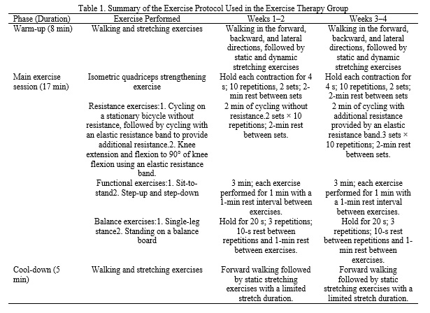
Participants in the exercise therapy group completed a 4-week resistance training program consisting of three sessions per week. In the present study, the resistance training program was designed in accordance with the fundamental principles of resistance training program design and based on the available literature in this field (22). The program was implemented after review and approval by experts. A detailed description of the exercise program and its implementation is presented in Table 1. The exercise program was performed for 4 weeks, with three sessions per week, each lasting 30 minutes. It is worth noting that all components of each training session, including warm-up and cool-down, were performed appropriately throughout the intervention period. Participants in the Orthokine therapy group received intra-articular Orthokine injections twice weekly for 4 weeks.
Osteoarthritis (OA) is one of the most common joint disorders, with a considerably higher prevalence in the knee than in any other joint of the human body (1). Compared with osteoarthritis affecting other joints, knee osteoarthritis (KOA) is associated with greater disability and more pronounced clinical manifestations, making it a major global health challenge (2). KOA is a leading cause of pain and functional limitation across all segments of the population, particularly among individuals over 50 years of age. Its prevalence has doubled since the mid-twentieth century and is expected to rise at an even faster rate in the future (3). The mechanisms underlying OA development are complex. One hypothesis suggests that cartilage degradation and the subsequent release of cartilage debris into the synovial fluid trigger inflammatory responses. Another hypothesis proposes that microscopic fractures in the subchondral bone lead to sclerosis and hypertrophy, thereby contributing to cartilage wear (4). In both pathways, the surrounding muscles, tendons, and ligaments weaken and undergo physiological changes, which can result in pain, reduced range of motion, and joint swelling (1, 3). Consequently, patients tend to adopt a more sedentary lifestyle, leading to a substantial decline in their quality of life (3).
To date, no definitive cure for KOA has been established (5), and most currently available interventions have been developed to manage the disease by reducing pain, slowing cartilage degeneration, and improving joint function (1, 3, 6). Current management strategies for knee osteoarthritis include pharmacological and non-pharmacological treatments, stem cell therapy, and joint surgery. Based on the available evidence, pharmacological and non-pharmacological approaches, such as exercise therapy, weight reduction, and the use of orthotic devices, are generally recommended as first-line treatments for patients with knee osteoarthritis (6). Among these, exercise therapy is recognized as one of the cornerstones of conservative management and plays an important role in reducing pain, improving joint function, and controlling disease progression by enhancing muscle strength, improving proprioception, and optimizing neuromuscular responses (7). The National Institute for Health and Care Excellence (NICE) identifies exercise therapy as the primary non-pharmacological intervention for the management of knee osteoarthritis. Evidence indicates that physical activity can alleviate symptoms and improve functional outcomes in patients with knee osteoarthritis by modulating inflammatory and metabolic processes within the joint (8). The findings of several systematic reviews and meta-analyses have demonstrated that exercise therapy can significantly improve muscle function, increase joint range of motion, and reduce pain, thereby enhancing physical function and quality of life in patients with knee osteoarthritis (9). Nevertheless, the implementation of exercise therapy in the management of knee osteoarthritis remains limited or is often delivered with suboptimal quality due to factors such as the prolonged duration required for treatment and therapeutic effectiveness, the absence of a standardized and universally accepted treatment protocol for knee osteoarthritis, physicians’ limited adherence to completing the full course of treatment, and patients’ poor compliance with performing progressive exercise because of pain and fear of physical activity (6, 10).
Conversely, some researchers have reported that pharmacological treatments may be effective in alleviating symptoms, controlling inflammation, and delaying joint replacement surgery in patients with knee osteoarthritis. Pharmacological treatment is generally recommended when patients’ symptoms fail to improve after approximately 3 to 6 months of non-pharmacological management (11). Given the pivotal role of inflammation in osteoarthritis-related joint damage, many pharmacological approaches have been developed to inhibit inflammatory processes (12). Although anti-inflammatory medications can be effective in controlling pain and inflammation, concerns regarding their adverse effects, particularly in middle-aged and older adults, have led to increasing interest in local and intra-articular treatment approaches (13). In this context, intra-articular injections have emerged as a less invasive therapeutic option, among which Orthokine therapy (Autologous Conditioned Serum; ACS) is considered one of the available treatment modalities (10).
Orthokine therapy (Autologous Conditioned Serum; ACS) is a conservative, autologous blood-based treatment designed to suppress inflammatory processes associated with osteoarthritis. In this procedure, serum obtained from incubated autologous blood is centrifuged and subsequently administered via intra-articular injection (14). The development of this technique has provided a novel approach to controlling joint inflammation and managing osteoarthritis and has been proposed as a potential alternative to the long-term use of anti-inflammatory medications (12). Currently, many orthopedic surgeons have shown considerable interest in this treatment approach, to the extent that it may even be considered among the primary therapeutic options for patients with osteoarthritis. Several studies have investigated the effects of Orthokine therapy on knee osteoarthritis. Baltzer et al. (2009) reported significant reductions in pain and improvements in physical function following Orthokine therapy (14). Gudack et al. also reported that Orthokine therapy is a safe treatment for knee osteoarthritis, is not associated with age-related limitations, and provides more sustained therapeutic effects than other non-surgical treatment modalities (12). However, Zaringam et al., who followed patients with knee osteoarthritis for up to 10 years after Orthokine therapy, reported that, in addition to being associated with a high incidence of adverse effects, this treatment neither prevented nor delayed joint replacement surgery (15). These findings indicate inconsistent results and uncertainty regarding the clinical effectiveness of this treatment approach. Despite the increasing use of Orthokine therapy in clinical and rehabilitation settings, conclusive evidence regarding its superiority or precise role compared with other conservative treatment approaches remains lacking (1). Furthermore, given the absence of a definitive cure for knee osteoarthritis, there is an increasing need for studies that simultaneously investigate and compare different management approaches (1, 3). Nevertheless, to the best of the authors’ knowledge, no previous study has compared the effects of Orthokine therapy and exercise therapy on symptom improvement and the management of osteoarthritis. Therefore, the present study aimed to compare the effects of exercise therapy and Orthokine therapy on pain intensity, joint stiffness, and quadriceps muscle strength in middle-aged women with knee osteoarthritis.
2. Methods
The present study was an applied quasi-experimental study with a two-group pretest–posttest design. Twenty middle-aged women with knee osteoarthritis were recruited using a purposive, non-random sampling method. The participants were then randomly allocated to either the exercise therapy group or the Orthokine therapy group using a simple randomization procedure based on a random number table. This approach helped minimize allocation bias between the study groups. The sample size was determined based on previous studies conducted in this field (16, 17). The study protocol was reviewed and approved by the relevant ethics committee and was conducted in accordance with ethical principles for research involving human participants. The inclusion criteria were as follows: age between 40 and 60 years; diagnosis of knee osteoarthritis based on clinical and radiographic findings with Kellgren–Lawrence grades 1–3; absence of cardiovascular disease, immune system disorders, neurological disorders, inflammatory or infectious arthropathies; no history of surgery or intra-articular treatment within the previous 6 months; absence of severe obesity (12, 14, 18); ability to perform exercise independently; and absence of other knee disorders (14, 18).
Eligibility was determined by specialist physicians in the fields of orthopedic surgery, rheumatology, and physical medicine and rehabilitation. The occurrence of any injury, inflammation, or swelling during the study, exacerbation of participants' pain attributable to the therapeutic interventions, or unwillingness to continue participation in the study were considered the exclusion criteria. Following participant recruitment, a briefing session was held during which the study procedures were explained to all participants. All participants provided written informed consent by signing a consent form before the study commenced. All participants were fully informed about the study procedures, and their personal information was kept confidential.
The participants were randomly assigned to either the exercise therapy group (n = 10) or the Orthokine therapy group (n = 10). Before the therapeutic interventions began, all participants completed the WOMAC questionnaire and the Visual Analog Scale (VAS). Participants' medication and analgesic use were monitored and recorded by an orthopedic surgeon (one of the study authors), and body mass index (BMI) was measured before the intervention and reported in the participants' characteristics table. In addition, participants in both groups were instructed not to participate in any other exercise programs or physical activities so that the effects of the primary study interventions could be evaluated independently. The WOMAC questionnaire is an internationally recognized and standardized instrument for evaluating treatment outcomes in patients with knee osteoarthritis and consists of 24 items assessing three domains: pain (5 items), morning joint stiffness (2 items), and physical function (17 items). Each item is scored on a five-point scale ranging from 0 to 4, yielding a total score ranging from 0 to 100, with higher scores indicating greater pain, more severe morning joint stiffness, and poorer physical function (19). In the present study, the WOMAC questionnaire was used solely to quantify morning knee joint stiffness. Previous studies have demonstrated that the pain and joint stiffness subscales of the WOMAC questionnaire have acceptable validity and reliability in patients with knee osteoarthritis (20).
Pain intensity was assessed using the Visual Analog Scale (VAS), which has been reported to have high sensitivity and validity for quantifying pain intensity in individuals with knee osteoarthritis (19). The VAS consists of a 100-mm horizontal line, with the left end labeled "no pain" and the right end labeled "worst pain imaginable." Participants were asked the general question, "How severe was your pain during the previous week?" They then indicated their pain intensity by marking a point on the line corresponding to a value between 0 and 100, where 0 represented no pain, and 100 represented the worst pain imaginable (19). Quadriceps muscle strength was measured using a handheld dynamometer by the same trained examiner during both the pretest and posttest assessments. In this procedure, one end of the dynamometer was secured to the participant's ankle, approximately 5 cm above the lateral malleolus, while the other end was fixed to a wall. The participant was seated on a chair and performed a knee extension, and the value displayed on the dynamometer was recorded as quadriceps muscle strength. The test was repeated three times for each participant, and the mean of the three measurements was used for the statistical analysis (21).
Participants in the exercise therapy group completed a 4-week resistance training program consisting of three sessions per week. In the present study, the resistance training program was designed in accordance with the fundamental principles of resistance training program design and based on the available literature in this field (22). The program was implemented after review and approval by experts. A detailed description of the exercise program and its implementation is presented in Table 1. The exercise program was performed for 4 weeks, with three sessions per week, each lasting 30 minutes. It is worth noting that all components of each training session, including warm-up and cool-down, were performed appropriately throughout the intervention period. Participants in the Orthokine therapy group received intra-articular Orthokine injections twice weekly for 4 weeks.
Orthokine was prepared as follows. First, 50–60 mL of venous blood was collected from each participant under sterile conditions using specialized syringes containing spherical glass beads. These glass beads stimulate the production of interleukin-1 receptor antagonist (IL-1Ra) by white blood cells during incubation (14, 23). The syringe containing the participant's blood was incubated for 24 h at 37°C in an atmosphere containing 5% carbon dioxide. After 24 h, the blood sample was centrifuged for 10 min. Subsequently, 10 mL of the serum-containing supernatant was filtered, aliquoted into sterile 2-mL microtubes, and stored at −20°C. In addition, before administration to the respective participant, the serum was tested for human immunodeficiency virus (HIV), syphilis, hepatitis B, and hepatitis C (23). For intra-articular Orthokine injection, the injection site was first prepared under sterile conditions. A 21-gauge needle was then inserted into the joint through the anterolateral approach, and a small amount of synovial fluid was aspirated to minimize dilution of the injected preparation. While the needle remained in place, 2 mL of Orthokine was injected into the joint. The injections were administered twice weekly for 4 weeks. Post-injection care instructions were provided to all participants (14).
Following completion of the therapeutic interventions, pain intensity, morning joint stiffness, and quadriceps muscle strength were reassessed. Data normality was evaluated using the Shapiro–Wilk test, and the results indicated that all study variables were normally distributed. In addition, the homogeneity of variances was confirmed using Levene's test. As all underlying assumptions were satisfied, parametric statistical tests were used for data analysis. An independent-samples t-test was used to compare the participants' demographic characteristics. To evaluate changes in the dependent variables over time and compare the patterns of these changes between the two groups in the pretest–posttest design, repeated-measures analysis of variance (ANOVA) was performed using SPSS software (version 23; SPSS Inc., Chicago, IL), with the level of statistical significance set at p ≤ 0.05.
3. Results
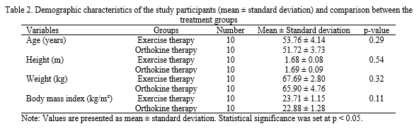
The demographic characteristics of the participants are presented in Table 2. As shown, there were no significant differences in demographic characteristics between the exercise therapy and orthokine therapy groups.
Following completion of the therapeutic interventions, pain intensity, morning joint stiffness, and quadriceps muscle strength were reassessed. Data normality was evaluated using the Shapiro–Wilk test, and the results indicated that all study variables were normally distributed. In addition, the homogeneity of variances was confirmed using Levene's test. As all underlying assumptions were satisfied, parametric statistical tests were used for data analysis. An independent-samples t-test was used to compare the participants' demographic characteristics. To evaluate changes in the dependent variables over time and compare the patterns of these changes between the two groups in the pretest–posttest design, repeated-measures analysis of variance (ANOVA) was performed using SPSS software (version 23; SPSS Inc., Chicago, IL), with the level of statistical significance set at p ≤ 0.05.
3. Results
The demographic characteristics of the participants are presented in Table 2. As shown, there were no significant differences in demographic characteristics between the exercise therapy and orthokine therapy groups.
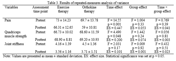
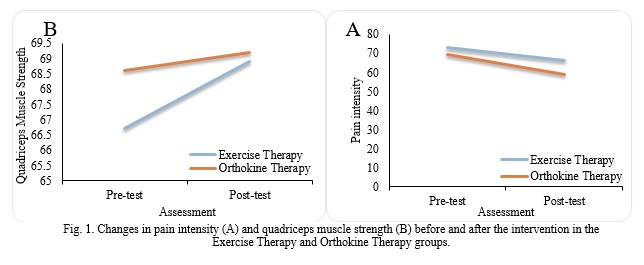
The findings obtained from repeated-measures analysis of variance were used to examine the effects of time (pretest to posttest), group, and the time × group interaction on pain intensity, joint stiffness, and quadriceps muscle strength, and are presented in Table 3. The results regarding pain intensity showed that the time effect was significant (F = 14.55, p = 0.001), indicating a significant reduction in pain intensity from pretest to posttest in both the exercise therapy and orthokine therapy groups. The reported effect size for pain intensity was large (ES = 0.447), demonstrating that this reduction was also clinically meaningful. On average, pain intensity in the exercise therapy group decreased from 73 ± 14.23 at pretest to 66.31 ± 12.65 at posttest, representing a 9.16% reduction. In the orthokine therapy group, pain intensity decreased from 69.7 ± 13.78 at pretest to 59 ± 10.81 at posttest, indicating a 15.3% reduction from pretest to posttest (Fig. 1).
In contrast, the group effect on pain intensity was not significant (F = 1.004, p = 0.33) and had a small effect size (ES = 0.053). Moreover, the time × group interaction effect was not significant (F = 0.769, p = 0.39) and was associated with a small effect size (ES = 0.041), indicating that the pattern of reduction in pain intensity from pretest to posttest was similar between the two groups.
Regarding knee joint stiffness, the results showed that the time effect was not significant (F = 2.031, p = 0.17), indicating no significant change in joint stiffness intensity from pretest to posttest in either group. The effect size was small (ES = 0.101). The group effect on joint stiffness intensity was also not significant (F = 0.009, p = 0.92), with a very small effect size (ES = 0.001). Furthermore, the time × group interaction did not reach statistical significance (F = 0.432, p = 0.51) and had a small effect size (ES = 0.023), indicating that the pattern of changes in joint stiffness from pretest to posttest was similar between the exercise therapy and orthokine therapy groups. These findings indicate that neither intervention had a significant or clinically meaningful effect on knee joint stiffness intensity. Regarding quadriceps muscle strength, the results of repeated-measures analysis of variance showed that the time effect was significant (F = 4.490, p = 0.048), indicating a significant increase in quadriceps muscle strength from pretest to posttest in both groups. The effect size was moderate (ES = 0.200). The mean quadriceps muscle strength in the exercise therapy group increased from 66.73 ± 10.02 at pretest to 68.90 ± 8.81 at posttest, representing an approximately 3.2% increase. In the orthokine therapy group, this value increased from 68.60 ± 11.59 at pretest to 69.20 ± 10.93 at posttest, representing a 0.8% increase compared with pretest values (Fig. 1). However, the group effect on quadriceps muscle strength was not significant (F = 1.442, p = 0.24), and the effect size was small (ES = 0.074). Moreover, the time × group interaction was not significant (F = 0.056, p = 0.81) and was associated with a very small effect size (ES = 0.003), indicating that the pattern of increase in quadriceps muscle strength from pretest to posttest was similar between the two groups. Overall, both interventions resulted in a significant improvement in quadriceps muscle strength; however, no significant difference was observed in the magnitude of changes between the two groups. Changes in the mean pain intensity and quadriceps muscle strength in the exercise therapy and Orthokine therapy groups from pretest to posttest are presented in Fig. 1.
4. Discussion
The main objective of the present study was to compare the effects of two approaches, exercise therapy and Orthokine therapy, on pain intensity, quadriceps muscle strength, and knee joint stiffness intensity in middle-aged women with knee osteoarthritis. The results showed that both interventions led to significant reductions in pain intensity and significant increases in quadriceps muscle strength from pretest to posttest, whereas neither intervention had a significant effect on knee joint stiffness intensity. Furthermore, no significant differences were observed between the two groups across the variables examined, indicating that exercise therapy and Orthokine therapy are similarly effective for these outcomes. In addition to statistical significance, the findings related to effect sizes and percentage changes indicated that both interventions produced clinically meaningful improvements in reducing pain and increasing muscle strength. Since achieving effective therapeutic approaches with minimal adverse effects is one of the primary goals of medicine and rehabilitation in conditions such as osteoarthritis, the findings of this study may provide valuable information for clinical decision-making regarding the selection of non-invasive interventions.
Regarding exercise therapy, the findings of the present study are consistent with previous studies reporting improvements in pain, muscle strength, and quality of life in patients with knee osteoarthritis (3, 24–27). From a mechanical perspective, exercise therapy improves knee joint stability and reduces the load imposed on joint structures by strengthening the muscles surrounding the joint, particularly the quadriceps muscles, which may ultimately lead to pain reduction (28, 29). Quadriceps muscle weakness is not only a risk factor for the development of knee osteoarthritis but also an important factor contributing to the progression of this condition (30, 31). The mechanism underlying quadriceps muscle weakness in knee osteoarthritis has been attributed to impairments in the neuromuscular function of these muscles, including arthrogenic muscle inhibition and a reduced ability to achieve complete voluntary muscle activation (21). Quadriceps muscle weakness reduces the capacity to absorb and distribute forces applied to the knee joint during daily activities. Consequently, greater mechanical loads are transferred to the passive structures of the joint, which may lead to microdamage in the subchondral bone and increased bone remodeling and sclerosis. These changes are accompanied by reduced shock-absorbing capacity of the subchondral region and ultimately increase the stresses applied to the articular cartilage, accelerating the process of cartilage thinning and degradation. The accumulation of these alterations results in reduced effective knee joint function and exacerbation of osteoarthritis progression (25). Therefore, resistance exercises, by reducing mechanical joint loading and improving muscle function, may contribute to pain reduction and improved functional status in patients (25, 26).
A significant reduction in pain intensity was also observed in the Orthokine therapy group, which is consistent with the findings of Baltzer et al. (2009), Fox et al. (2010), and Barto et al. (2017) (14, 32, 33). Osteoarthritis is associated with increased levels of inflammatory cytokines, particularly interleukin-1, which plays a key role in cartilage degradation and pain development (34, 35). Previous studies have demonstrated that interleukin-1 levels are significantly elevated in patients with osteoarthritis and, through inhibition of type II collagen synthesis and reduction of aggrecan production, are considered one of the major contributors to articular cartilage degradation (35). Research evidence also indicates an association between interleukin-1 and pain severity in knee osteoarthritis. Therefore, interventions that reduce the levels of this cytokine may be effective in managing disease symptoms (13, 14). Orthokine therapy is one of the novel therapeutic approaches based on reducing inflammatory cytokines, particularly interleukin-1, and may play a role in improving symptoms and modulating the progression of knee osteoarthritis by reducing inflammation and enhancing reparative processes (34, 36). One of the advantages of this method is that the serum is prepared from the individual's own blood and injected directly into the affected joint, thereby increasing its biological safety. Accordingly, some researchers have considered Orthokine therapy a more effective option than pharmacological treatment and surgery for the management of knee osteoarthritis (36). However, the findings of the present study are inconsistent with those of Rutgers et al., who reported that Orthokine therapy did not have a significant effect on improving symptoms, particularly pain intensity, and introduced pharmacological treatment as a more appropriate option (37). The inconsistency between the findings of the present study and those of Rutgers et al. may be attributed to differences in the injected Orthokine dosage and the method used to prepare the injected serum.
The findings of the present study demonstrated that Orthokine therapy, in addition to reducing pain intensity, improved quadriceps muscle strength in patients with knee osteoarthritis, which is consistent with previous research findings, including those of Vitali et al. (2020) (36). One possible explanation for this finding is the bidirectional relationship between knee joint pain and the strength of the surrounding muscles. Accordingly, quadriceps muscle weakness is not only recognized as a risk factor for the development and progression of osteoarthritis, but evidence also indicates that the presence of pain can lead to reduced quadriceps and hamstring muscle strength (38). Joint pain is often accompanied by inflammation, swelling, and reduced range of motion, which can impair knee function (38). On the other hand, studies have reported that increased inflammatory cytokines are associated with reduced knee function and range of motion (39), and elevated levels of interleukin-1 may contribute to muscle atrophy and reduced muscle strength (40). Therefore, Orthokine therapy, through reducing inflammatory cytokines, particularly interleukin-1, may not only reduce pain but also potentially improve quadriceps muscle strength by decreasing joint inflammation and inhibiting muscle catabolic processes. Based on a review of the previous literature, no study was identified that had simultaneously and directly compared these two approaches, exercise therapy and Orthokine therapy, in patients with knee osteoarthritis. Therefore, the results of each intervention were compared separately with those of previous studies.
Most studies in the field of exercise therapy have focused on comparing different exercise protocols, and few studies have investigated the effects of exercise therapy compared with other therapeutic approaches, such as pharmacological treatment or intra-articular injections. In this regard, Thomas et al. (2023) reported that exercise therapy had a greater effect than collagen supplementation on improving range of motion and increasing thigh muscle strength in patients with knee osteoarthritis, whereas no significant difference was observed between the two approaches regarding pain intensity. However, the combination of exercise therapy and collagen supplementation resulted in greater improvement in symptoms (41). Kawasaki et al. (2009), by comparing home-based exercise therapy with intra-articular hyaluronic acid injection, demonstrated that both approaches had approximately similar effects on improving symptoms, including pain (42). Furthermore, Bandak et al. reported that eight weeks of exercise therapy combined with education had similar effectiveness to saline injection (placebo) in reducing pain and improving function in patients with knee osteoarthritis (43). The results of these studies are consistent with the findings of the present study.
In comparison, the role of Orthokine therapy in the management of osteoarthritis has been less extensively investigated. Baselga Garcia-Escudero et al., in a prospective study, demonstrated that the combination of Orthokine therapy and physiotherapy resulted in sustained improvements in pain, joint stiffness, and patient function for up to two years after treatment (44). Furthermore, Shirokova et al. reported that Orthokine therapy resulted in greater improvement in patients' symptoms compared with platelet-rich plasma (PRP) injection and was associated with fewer adverse effects (45). The differences between the findings of these studies and those of the present study may be attributed to the nature of the compared interventions, as Orthokine therapy was compared with other injection-based approaches in the aforementioned studies, whereas in the present study, this approach was compared with exercise therapy as a completely non-invasive intervention. Overall, considering the similar effectiveness of the two approaches in improving symptoms in patients with knee osteoarthritis, and taking into account the non-invasive nature, safety, easy accessibility, and cost-effectiveness of exercise therapy, the findings of the present study support the role of exercise therapy as a first-line therapeutic option in the management of knee osteoarthritis. However, achieving optimal outcomes requires the design of an appropriate exercise protocol and the adherence of both patients and therapists to the correct and complete implementation of the therapeutic program.
5. Conclusion
Based on the findings of the present study, exercise therapy and Orthokine therapy demonstrated approximately similar effects on reducing pain intensity and increasing quadriceps muscle strength in middle-aged individuals with knee osteoarthritis. Considering the similar effectiveness of the two approaches and the non-invasive, safe, and cost-effective nature of exercise therapy, the findings of the present study support the use of exercise therapy as an effective option for the management of knee osteoarthritis. However, further studies with larger sample sizes and longer follow-up periods are needed to confirm and generalize these findings.
Acknowledgments
The authors would like to express their gratitude to the medical centers and the Faculty of Medical Sciences of Mashhad for their support of this research. The authors also appreciate and thank all individuals who participated in this study.
Ethical Considerations
Compliance with ethical guidelines
This study was conducted in accordance with research ethics principles, and the study protocol was approved by the relevant ethics committee under the code (IR-KHU.KRC.1000.210). Written informed consent was obtained from all participants in accordance with these ethical guidelines.
Funding
The authors received no specific financial support for this research from any funding agency in the public, commercial, or not-for-profit sectors.
Authors' contributions
The first author (corresponding author) and the second author contributed to the study design and supervision, data collection, data analysis, and interpretation of the results. The third author contributed to the methodology, data analysis, and interpretation of the results. The fourth author contributed to data collection and the evaluation and examination of participants regarding the study inclusion and exclusion criteria. Finally, the fifth author contributed to the interpretation of the results, manuscript preparation and revision, as well as finalizing the manuscript.
Conflicts of interest
The authors declare that they have no conflict of interest associated with this study.
Regarding knee joint stiffness, the results showed that the time effect was not significant (F = 2.031, p = 0.17), indicating no significant change in joint stiffness intensity from pretest to posttest in either group. The effect size was small (ES = 0.101). The group effect on joint stiffness intensity was also not significant (F = 0.009, p = 0.92), with a very small effect size (ES = 0.001). Furthermore, the time × group interaction did not reach statistical significance (F = 0.432, p = 0.51) and had a small effect size (ES = 0.023), indicating that the pattern of changes in joint stiffness from pretest to posttest was similar between the exercise therapy and orthokine therapy groups. These findings indicate that neither intervention had a significant or clinically meaningful effect on knee joint stiffness intensity. Regarding quadriceps muscle strength, the results of repeated-measures analysis of variance showed that the time effect was significant (F = 4.490, p = 0.048), indicating a significant increase in quadriceps muscle strength from pretest to posttest in both groups. The effect size was moderate (ES = 0.200). The mean quadriceps muscle strength in the exercise therapy group increased from 66.73 ± 10.02 at pretest to 68.90 ± 8.81 at posttest, representing an approximately 3.2% increase. In the orthokine therapy group, this value increased from 68.60 ± 11.59 at pretest to 69.20 ± 10.93 at posttest, representing a 0.8% increase compared with pretest values (Fig. 1). However, the group effect on quadriceps muscle strength was not significant (F = 1.442, p = 0.24), and the effect size was small (ES = 0.074). Moreover, the time × group interaction was not significant (F = 0.056, p = 0.81) and was associated with a very small effect size (ES = 0.003), indicating that the pattern of increase in quadriceps muscle strength from pretest to posttest was similar between the two groups. Overall, both interventions resulted in a significant improvement in quadriceps muscle strength; however, no significant difference was observed in the magnitude of changes between the two groups. Changes in the mean pain intensity and quadriceps muscle strength in the exercise therapy and Orthokine therapy groups from pretest to posttest are presented in Fig. 1.
4. Discussion
The main objective of the present study was to compare the effects of two approaches, exercise therapy and Orthokine therapy, on pain intensity, quadriceps muscle strength, and knee joint stiffness intensity in middle-aged women with knee osteoarthritis. The results showed that both interventions led to significant reductions in pain intensity and significant increases in quadriceps muscle strength from pretest to posttest, whereas neither intervention had a significant effect on knee joint stiffness intensity. Furthermore, no significant differences were observed between the two groups across the variables examined, indicating that exercise therapy and Orthokine therapy are similarly effective for these outcomes. In addition to statistical significance, the findings related to effect sizes and percentage changes indicated that both interventions produced clinically meaningful improvements in reducing pain and increasing muscle strength. Since achieving effective therapeutic approaches with minimal adverse effects is one of the primary goals of medicine and rehabilitation in conditions such as osteoarthritis, the findings of this study may provide valuable information for clinical decision-making regarding the selection of non-invasive interventions.
Regarding exercise therapy, the findings of the present study are consistent with previous studies reporting improvements in pain, muscle strength, and quality of life in patients with knee osteoarthritis (3, 24–27). From a mechanical perspective, exercise therapy improves knee joint stability and reduces the load imposed on joint structures by strengthening the muscles surrounding the joint, particularly the quadriceps muscles, which may ultimately lead to pain reduction (28, 29). Quadriceps muscle weakness is not only a risk factor for the development of knee osteoarthritis but also an important factor contributing to the progression of this condition (30, 31). The mechanism underlying quadriceps muscle weakness in knee osteoarthritis has been attributed to impairments in the neuromuscular function of these muscles, including arthrogenic muscle inhibition and a reduced ability to achieve complete voluntary muscle activation (21). Quadriceps muscle weakness reduces the capacity to absorb and distribute forces applied to the knee joint during daily activities. Consequently, greater mechanical loads are transferred to the passive structures of the joint, which may lead to microdamage in the subchondral bone and increased bone remodeling and sclerosis. These changes are accompanied by reduced shock-absorbing capacity of the subchondral region and ultimately increase the stresses applied to the articular cartilage, accelerating the process of cartilage thinning and degradation. The accumulation of these alterations results in reduced effective knee joint function and exacerbation of osteoarthritis progression (25). Therefore, resistance exercises, by reducing mechanical joint loading and improving muscle function, may contribute to pain reduction and improved functional status in patients (25, 26).
A significant reduction in pain intensity was also observed in the Orthokine therapy group, which is consistent with the findings of Baltzer et al. (2009), Fox et al. (2010), and Barto et al. (2017) (14, 32, 33). Osteoarthritis is associated with increased levels of inflammatory cytokines, particularly interleukin-1, which plays a key role in cartilage degradation and pain development (34, 35). Previous studies have demonstrated that interleukin-1 levels are significantly elevated in patients with osteoarthritis and, through inhibition of type II collagen synthesis and reduction of aggrecan production, are considered one of the major contributors to articular cartilage degradation (35). Research evidence also indicates an association between interleukin-1 and pain severity in knee osteoarthritis. Therefore, interventions that reduce the levels of this cytokine may be effective in managing disease symptoms (13, 14). Orthokine therapy is one of the novel therapeutic approaches based on reducing inflammatory cytokines, particularly interleukin-1, and may play a role in improving symptoms and modulating the progression of knee osteoarthritis by reducing inflammation and enhancing reparative processes (34, 36). One of the advantages of this method is that the serum is prepared from the individual's own blood and injected directly into the affected joint, thereby increasing its biological safety. Accordingly, some researchers have considered Orthokine therapy a more effective option than pharmacological treatment and surgery for the management of knee osteoarthritis (36). However, the findings of the present study are inconsistent with those of Rutgers et al., who reported that Orthokine therapy did not have a significant effect on improving symptoms, particularly pain intensity, and introduced pharmacological treatment as a more appropriate option (37). The inconsistency between the findings of the present study and those of Rutgers et al. may be attributed to differences in the injected Orthokine dosage and the method used to prepare the injected serum.
The findings of the present study demonstrated that Orthokine therapy, in addition to reducing pain intensity, improved quadriceps muscle strength in patients with knee osteoarthritis, which is consistent with previous research findings, including those of Vitali et al. (2020) (36). One possible explanation for this finding is the bidirectional relationship between knee joint pain and the strength of the surrounding muscles. Accordingly, quadriceps muscle weakness is not only recognized as a risk factor for the development and progression of osteoarthritis, but evidence also indicates that the presence of pain can lead to reduced quadriceps and hamstring muscle strength (38). Joint pain is often accompanied by inflammation, swelling, and reduced range of motion, which can impair knee function (38). On the other hand, studies have reported that increased inflammatory cytokines are associated with reduced knee function and range of motion (39), and elevated levels of interleukin-1 may contribute to muscle atrophy and reduced muscle strength (40). Therefore, Orthokine therapy, through reducing inflammatory cytokines, particularly interleukin-1, may not only reduce pain but also potentially improve quadriceps muscle strength by decreasing joint inflammation and inhibiting muscle catabolic processes. Based on a review of the previous literature, no study was identified that had simultaneously and directly compared these two approaches, exercise therapy and Orthokine therapy, in patients with knee osteoarthritis. Therefore, the results of each intervention were compared separately with those of previous studies.
Most studies in the field of exercise therapy have focused on comparing different exercise protocols, and few studies have investigated the effects of exercise therapy compared with other therapeutic approaches, such as pharmacological treatment or intra-articular injections. In this regard, Thomas et al. (2023) reported that exercise therapy had a greater effect than collagen supplementation on improving range of motion and increasing thigh muscle strength in patients with knee osteoarthritis, whereas no significant difference was observed between the two approaches regarding pain intensity. However, the combination of exercise therapy and collagen supplementation resulted in greater improvement in symptoms (41). Kawasaki et al. (2009), by comparing home-based exercise therapy with intra-articular hyaluronic acid injection, demonstrated that both approaches had approximately similar effects on improving symptoms, including pain (42). Furthermore, Bandak et al. reported that eight weeks of exercise therapy combined with education had similar effectiveness to saline injection (placebo) in reducing pain and improving function in patients with knee osteoarthritis (43). The results of these studies are consistent with the findings of the present study.
In comparison, the role of Orthokine therapy in the management of osteoarthritis has been less extensively investigated. Baselga Garcia-Escudero et al., in a prospective study, demonstrated that the combination of Orthokine therapy and physiotherapy resulted in sustained improvements in pain, joint stiffness, and patient function for up to two years after treatment (44). Furthermore, Shirokova et al. reported that Orthokine therapy resulted in greater improvement in patients' symptoms compared with platelet-rich plasma (PRP) injection and was associated with fewer adverse effects (45). The differences between the findings of these studies and those of the present study may be attributed to the nature of the compared interventions, as Orthokine therapy was compared with other injection-based approaches in the aforementioned studies, whereas in the present study, this approach was compared with exercise therapy as a completely non-invasive intervention. Overall, considering the similar effectiveness of the two approaches in improving symptoms in patients with knee osteoarthritis, and taking into account the non-invasive nature, safety, easy accessibility, and cost-effectiveness of exercise therapy, the findings of the present study support the role of exercise therapy as a first-line therapeutic option in the management of knee osteoarthritis. However, achieving optimal outcomes requires the design of an appropriate exercise protocol and the adherence of both patients and therapists to the correct and complete implementation of the therapeutic program.
5. Conclusion
Based on the findings of the present study, exercise therapy and Orthokine therapy demonstrated approximately similar effects on reducing pain intensity and increasing quadriceps muscle strength in middle-aged individuals with knee osteoarthritis. Considering the similar effectiveness of the two approaches and the non-invasive, safe, and cost-effective nature of exercise therapy, the findings of the present study support the use of exercise therapy as an effective option for the management of knee osteoarthritis. However, further studies with larger sample sizes and longer follow-up periods are needed to confirm and generalize these findings.
Acknowledgments
The authors would like to express their gratitude to the medical centers and the Faculty of Medical Sciences of Mashhad for their support of this research. The authors also appreciate and thank all individuals who participated in this study.
Ethical Considerations
Compliance with ethical guidelines
This study was conducted in accordance with research ethics principles, and the study protocol was approved by the relevant ethics committee under the code (IR-KHU.KRC.1000.210). Written informed consent was obtained from all participants in accordance with these ethical guidelines.
Funding
The authors received no specific financial support for this research from any funding agency in the public, commercial, or not-for-profit sectors.
Authors' contributions
The first author (corresponding author) and the second author contributed to the study design and supervision, data collection, data analysis, and interpretation of the results. The third author contributed to the methodology, data analysis, and interpretation of the results. The fourth author contributed to data collection and the evaluation and examination of participants regarding the study inclusion and exclusion criteria. Finally, the fifth author contributed to the interpretation of the results, manuscript preparation and revision, as well as finalizing the manuscript.
Conflicts of interest
The authors declare that they have no conflict of interest associated with this study.
Type of Study: Applicable |
Subject:
Special
Received: 2025/08/27 | Accepted: 2026/07/27 | Published: 2026/07/30
Received: 2025/08/27 | Accepted: 2026/07/27 | Published: 2026/07/30
References
1. Geng R, Li J, Yu C, Zhang C, Chen F, Chen J, et al. Knee osteoarthritis: current status and research progress in treatment. Experimental and Therapeutic Medicine. 2023;26(4):1-11. [DOI:10.3892/etm.2023.12180]
2. Alikhani M, Tabatabai H. The effects of six weeks of patella taping on function, proprioception, and balance in women with knee osteoarthritis. Journal of Sport Biomechanics. 2024;10(3):188-201. [DOI:10.61186/JSportBiomech.10.3.188]
3. Raposo F, Ramos M, Cruz AL. Effects of exercise on knee osteoarthritis: a systematic review. Musculoskeletal Care. 2021;19(4):399-435. [DOI:10.1002/msc.1538]
4. Berenbaum F. Osteoarthritis as an inflammatory disease (osteoarthritis is not osteoarthrosis!). Osteoarthritis and Cartilage. 2013;21(1):16-21. [DOI:10.1016/j.joca.2012.11.012]
5. Bavardi Moghadam E, Shojaedin SS. The effect of a period of stretching training on functional dynamic balance performance and range of motion in patients with knee osteoarthritis. Journal of Sport Biomechanics. 2017;2(4):5-18.
6. Bell E, Wallis J, Goff AJ, Crossley K, O'Halloran P, Barton C. Does land-based exercise therapy improve physical activity in people with knee osteoarthritis? A systematic review with meta-analyses. Osteoarthritis and Cartilage. 2022;30(11):1420-1433. [DOI:10.1016/j.joca.2022.07.008]
7. Kiani Dooghabadi F, Khoshraftar Yazdi N, Farzaneh H. The effect of a period of kinesiotherapy and reflexology on pain intensity and range of motion in elderly women with knee osteoarthritis. Journal of Sport Biomechanics. 2020;6(2):98-109. [DOI:10.32598/biomechanics.6.2.2]
8. Leong D, Sun H. Osteoarthritis: why exercise? Journal of Exercise and Sports Orthopedics. 2013;1(1):04. [DOI:10.15226/2374-6904/1/1/00104]
9. Mo L, Jiang B, Mei T, Zhou D. Exercise therapy for knee osteoarthritis: a systematic review and network meta-analysis. Orthopaedic Journal of Sports Medicine. 2023;11(5):23259671231172773. [DOI:10.1177/23259671231172773]
10. Nelligan RK, Hinman RS, Kasza J, Crofts SJ, Bennell KL. Effects of a self-directed web-based strengthening exercise and physical activity program supported by automated text messages for people with knee osteoarthritis: a randomized clinical trial. JAMA Internal Medicine. 2021;181(6):776-785. [DOI:10.1001/jamainternmed.2021.0991]
11. Arden NK, Perry TA, Bannuru RR, Bruyère O, Cooper C, Haugen IK, et al. Non-surgical management of knee osteoarthritis: comparison of ESCEO and OARSI 2019 guidelines. Nature Reviews Rheumatology. 2021;17(1):59-66. [DOI:10.1038/s41584-020-00523-9]
12. Godek P, Szajkowski S, Golicki D. Evaluation of the effectiveness of Orthokine therapy: retrospective analysis of 1000 cases. Ortopedia Traumatologia Rehabilitacja. 2020;22(2):107-119. [DOI:10.5604/01.3001.0014.1167]
13. Schjerning Olsen AM, Fosbøl EL, Lindhardsen J, Folke F, Charlot M, Selmer C, et al. Duration of treatment with nonsteroidal anti-inflammatory drugs and impact on risk of death and recurrent myocardial infarction in patients with prior myocardial infarction: a nationwide cohort study. Circulation. 2011;123(20):2226-2235. [DOI:10.1161/CIRCULATIONAHA.110.004671]
14. Baltzer A, Moser C, Jansen S, Krauspe R. Autologous conditioned serum (Orthokine) is an effective treatment for knee osteoarthritis. Osteoarthritis and Cartilage. 2009;17(2):152-160. [DOI:10.1016/j.joca.2008.06.014]
15. Zarringam D, Bekkers JE, Saris DB. Long-term effect of injection treatment for osteoarthritis in the knee by Orthokine autologous conditioned serum. Cartilage. 2018;9(2):140-145. [DOI:10.1177/1947603517743001]
16. Piran Hamlabadi M, Jafarnezhadgero AA, Noorian E. The effect of 8 sessions of balance exercises on proprioception of knee and ankle joints in diabetic patients with knee osteoarthritis. Journal of Anesthesia and Pain. 2021;12(3):50-59.
17. Mazloum V, Sobhani V, Shirvani H, Khatibi AA. A comparative study on the influence of Kinesio Taping and laser therapy on knee joint position sense, pain intensity, and function in individuals with knee osteoarthritis. Journal of Shahrekord University of Medical Sciences. 2016; 18(5): 103-114.
18. Yang KGA, Raijmakers NJ, Van Arkel ERA, Caron JJ, Rijk PC, Willems WJ, et al. Autologous interleukin-1 receptor antagonist improves function and symptoms in osteoarthritis when compared to placebo in a prospective randomized controlled trial. Osteoarthritis and Cartilage. 2008;16(4):498-505. [DOI:10.1016/j.joca.2007.07.008]
19. Barati S, Khayambashi K, Rahnama N, Nayeri M. Effect of a selected core stabilization training program on pain and function in females with knee osteoarthritis. Journal of Research in Rehabilitation Sciences. 2012;8(1):40-48.
20. Ansari G, Delbari A, Karimi M, Akbari Kamrani AA, Mohammadi S, Sahaf R. Effect of Melilotus officinalis oil on knee joint pain and stiffness in the elderly with primary knee osteoarthritis. Salmand: Iranian Journal of Ageing. 2020;15(2):160-175. [DOI:10.32598/sija.13.10.520]
21. Heidari B, Javadian Y, Akbari R. Quadriceps muscle strength in patients with knee osteoarthritis. Journal of Advances in Medical and Biomedical Research. 2016;24(102):111-120.
22. Ezadpanah A, Moazami M, Khoshraftar Yazdi N. Effect of a period of therapeutic exercise and detraining on balance in women with knee osteoarthritis. Journal of Modern Rehabilitation. 2016;9(5):101-109.
23. Baltzer AW, Ostapczuk MS, Stosch D, Seidel F, Granrath M. A new treatment for hip osteoarthritis: clinical evidence for the efficacy of autologous conditioned serum. Orthopedic Reviews. 2013;5(2):e13. [DOI:10.4081/or.2013.e13]
24. Al-Khlaifat L, Herrington LC, Tyson SF, Hammond A, Jones RK. The effectiveness of an exercise programme on dynamic balance in patients with medial knee osteoarthritis: a pilot study. The Knee. 2016;23(5):849-856. [DOI:10.1016/j.knee.2016.05.006]
25. Miri H, Tabatabaeineghad M, Sharifi S, Ardakanizadeh M, Rangraz E. The influence of eight weeks of resistance training with Triband on motor function, pain severity, and strength in elderly women with knee osteoarthritis. Journal of Sports and Biomotor Sciences. 2023;15(29):108-116.
26. Patterson BE, Girdwood MA, West TJ, Bruder AM, Øiestad BE, Juhl C, et al. Muscle strength and osteoarthritis of the knee: a systematic review and meta-analysis of longitudinal studies. Skeletal Radiology. 2023;52(11):2085-2097. [DOI:10.1007/s00256-022-04266-4]
27. Prabhakar AJ, Shruthi R, Thomas DT, Nayak P, Joshua AM, Prabhu S, et al. Effectiveness of balance training on pain and functional outcomes in knee osteoarthritis: a systematic review and meta-analysis. F1000Research. 2023;11:598. [DOI:10.12688/f1000research.111998.2]
28. Rice DA, McNair PJ, Lewis GN. Mechanisms of quadriceps muscle weakness in knee joint osteoarthritis: the effects of prolonged vibration on torque and muscle activation in osteoarthritic and healthy control subjects. Arthritis Research & Therapy. 2011;13:R151. [DOI:10.1186/ar3467]
29. Zeng CY, Zhang ZR, Tang ZM, Hua FZ. Benefits and mechanisms of exercise training for knee osteoarthritis. Frontiers in Physiology. 2021;12:794062. [DOI:10.3389/fphys.2021.794062]
30. Alshahrani MS, Reddy RS. Quadriceps strength, postural stability, and pain mediation in bilateral knee osteoarthritis: a comparative analysis with healthy controls. Diagnostics. 2023;13(19):3110. [DOI:10.3390/diagnostics13193110]
31. Øiestad BE, Juhl CB, Culvenor AG, Berg B, Thorlund JB. Knee extensor muscle weakness is a risk factor for the development of knee osteoarthritis: an updated systematic review and meta-analysis including 46,819 men and women. British Journal of Sports Medicine. 2022;56(6):349-355. [DOI:10.1136/bjsports-2021-104861]
32. Barreto A, Braun TR. A new treatment for knee osteoarthritis: clinical evidence for the efficacy of Arthrokinex autologous conditioned serum. Journal of Orthopaedics. 2017;14(1):4-9. [DOI:10.1016/j.jor.2016.10.008]
33. Fox BA, Stephens MM. Treatment of knee osteoarthritis with Orthokine-derived autologous conditioned serum. Expert Review of Clinical Immunology. 2010;6(3):335-345. [DOI:10.1586/eci.10.17]
34. Kılınç BE, Öç Y. Evaluation of autologous conditioned serum in the treatment of osteoarthritis. Archives of Clinical and Experimental Medicine. 2019;4(2):94-98. [DOI:10.25000/acem.569936]
35. Motaal FKAE, Elganzoury AM, Fathalla MM, Abdulkareem O. Low-dose intra-articular autologous conditioned serum in the treatment of primary knee osteoarthritis. Egyptian Rheumatology and Rehabilitation. 2014;41:98-102. [DOI:10.4103/1110-161X.140523]
36. Vitali M, Ometti M, Drossinos A, Pironti P, Santoleri L, Salini V. Autologous conditioned serum: clinical and functional results using a novel disease-modifying agent for the management of knee osteoarthritis. Journal of Drug Assessment. 2020;9(1):43-51. [DOI:10.1080/21556660.2020.1734009]
37. Rutgers M, Creemers LB, Yang KGA, Raijmakers NJH, Dhert WJA, Saris DBF. Osteoarthritis treatment using autologous conditioned serum after placebo: patient considerations and clinical response in a non-randomized case series. Acta Orthopaedica. 2015;86(1):114-118. [DOI:10.3109/17453674.2014.950467]
38. Ruhdorfer A, Wirth W, Eckstein F. Association of knee pain with a reduction in thigh muscle strength: a cross-sectional analysis including 4553 Osteoarthritis Initiative participants. Osteoarthritis and Cartilage. 2017;25(5):658-666. [DOI:10.1016/j.joca.2016.10.026]
39. Nees TA, Rosshirt N, Zhang JA, Reiner T, Sorbi R, Tripel E, et al. Synovial cytokines significantly correlate with osteoarthritis-related knee pain and disability: inflammatory mediators of potential clinical relevance. Journal of Clinical Medicine. 2019;8(9):1343. [DOI:10.3390/jcm8091343]
40. Kanai M, Ganbaatar B, Endo I, Ohnishi Y, Teramachi J, Tenshin H, et al. Inflammatory cytokine-induced muscle atrophy and weakness can be ameliorated by inhibition of TGF-β-activated kinase 1. International Journal of Molecular Sciences. 2024;25(11):5715. [DOI:10.3390/ijms25115715]
41. Thomas DT, Prabhakar AJ, Eapen C, Patel VD, Palaniswamy V, Dsouza MC, et al. Comparison of single and combined treatment with exercise therapy and collagen supplementation on early knee arthritis among athletes: a quasi-randomized trial. International Journal of Environmental Research and Public Health. 2023;20(23):7088. [DOI:10.3390/ijerph20237088]
42. Kawasaki T, Kurosawa H, Ikeda H, Takazawa Y, Ishijima M, Kubota M, et al. Therapeutic home exercise versus intra-articular hyaluronate injection for osteoarthritis of the knee: a 6-month prospective randomized open-label trial. Journal of Orthopaedic Science. 2009;14:182-191. [DOI:10.1007/s00776-008-1312-9]
43. Bandak E, Christensen R, Overgaard A, Kristensen LE, Ellegaard K, Guldberg-Møller J, et al. Exercise and education versus saline injections for knee osteoarthritis: a randomized controlled equivalence trial. Annals of the Rheumatic Diseases. 2022;81(4):537-543. [DOI:10.1136/annrheumdis-2021-221129]
44. Baselga García-Escudero J, Miguel Hernández Trillos P. Treatment of osteoarthritis of the knee with a combination of autologous conditioned serum and physiotherapy: a two-year observational study. PLoS One. 2015;10(12):e0145551. [DOI:10.1371/journal.pone.0145551]
45. Shirokova L, Noskov S, Gorokhova V, Reinecke J, Shirokova K. Intra-articular injections of a whole blood clot secretome, autologous conditioned serum, have superior clinical and biochemical efficacy over platelet-rich plasma and induce rejuvenation-associated changes of joint metabolism: a prospective controlled open-label clinical study in chronic knee osteoarthritis. Rejuvenation Research. 2020;23(5):401-410. [DOI:10.1089/rej.2019.2263]
| Rights and permissions | |
 |
This work is licensed under a Creative Commons Attribution-NonCommercial 4.0 International License. |

Journal of Sport Biomechanics
Department of Sport Biomechanics, Faculty of Humanities, Islamic Azad University, Hamedan Branch, Prof. Mousivand Blvd, Imam Khomeini Blvd, Hamedan, Iran.
Journal Tel: +98 81 34494042
Website: http://biomechanics.iauh.ac.ir
Email: sportbiomechanics@iauh.ac.ir
