

Volume 12, Issue 4 (2-2027)
J Sport Biomech 2027, 12(4): 756-770 |
Back to browse issues page




Download citation:
BibTeX | RIS | EndNote | Medlars | ProCite | Reference Manager | RefWorks
Send citation to:


BibTeX | RIS | EndNote | Medlars | ProCite | Reference Manager | RefWorks
Send citation to:
Madadi-Shad M, Bagheri S, Mahdieh I, Ghasemi M. The Effect of Kinesiotaping of the Biceps Femoris and Vastus Lateralis Muscles on External Knee Adduction Moment and Lower-limb Joint Power in Individuals with Genu Varum and Healthy Controls During Walking. J Sport Biomech 2027; 12 (4) :756-770
URL: http://biomechanics.iauh.ac.ir/article-1-569-en.html
URL: http://biomechanics.iauh.ac.ir/article-1-569-en.html


1- Department of Physical Education, Farhangian University, P.O.Box 14665-889, Tehran, Iran.
2- Department of Biomedical Science, University of Padova, Padova, Italy.
2- Department of Biomedical Science, University of Padova, Padova, Italy.
Full-Text [PDF 1572 kb]
(8 Downloads)
| Abstract (HTML) (32 Views)
Full-Text: (7 Views)
Introduction
Genu varum is a lower-limb alignment deformity characterized by lateral bowing of the legs and increased separation between the knees when standing with the ankles together. Previous studies have identified genu varum as a potential risk factor for the progression of knee osteoarthritis (OA) (1). The prevalence of knee OA has been reported to be approximately 12% among adults aged 65 years and older (2). Furthermore, the risk of medial compartment knee OA, which is associated with genu varum, has been reported to be approximately 10 times higher than that of lateral compartment OA, which is associated with genu valgum (3). Individuals with genu varum typically exhibit an altered distribution of mechanical loads across the knee joint (4), potentially increasing the loading of the medial compartment and contributing to OA progression.
Among non-invasive interventions aimed at modifying knee joint loading, kinesiotaping has gained considerable attention (5). Kinesiotaping is commonly used to provide external support and potentially influence neuromuscular function and joint mechanics. However, its effects on knee joint loading and lower-limb biomechanics during walking remain insufficiently established, particularly in individuals with genu varum. Further investigation is therefore needed to determine whether kinesiotaping can meaningfully alter the mechanical demands placed on the knee and other lower-limb joints during gait.
The present study aimed to investigate the immediate effects of kinesiotaping applied to the biceps femoris and vastus lateralis muscles on the external knee adduction moment and lower-limb joint power during walking in individuals with genu varum and healthy controls. It was hypothesized that kinesiotaping would reduce the external knee adduction moment and increase lower-limb joint power in individuals with genu varum during walking.
Methods
Thirty male volunteers participated in this study, with a mean age of 21.64 ± 5.40 years, a mean height of 179.25 ± 6.12 cm, and a mean body mass of 73.15 ± 8.14 kg. Participants were divided into two groups of 15: individuals with genu varum and healthy controls. The control group included individuals with an intercondylar distance (measured between the medial femoral epicondyles) of less than 6 cm in the standing position (6) and a Q-angle ranging from 13° to 18° (7).
Gait kinematics were recorded using a Vicon three-dimensional motion capture system operating at a sampling frequency of 100 Hz, with Nexus software (version 1.7.5). Ground reaction forces were recorded at 1,000 Hz using two Kistler force plates embedded in the walkway. Participants performed walking trials under two conditions: without kinesiotaping and with kinesiotaping applied to the biceps femoris and vastus lateralis muscles.
A 2 × 2 mixed-design analysis of variance (ANOVA), with group (genu varum vs. control) as the between-subjects factor and taping condition (without vs. with kinesiotaping) as the within-subjects factor, was used to examine the main and interaction effects on the external knee adduction moment and lower-limb joint power (7).
Results
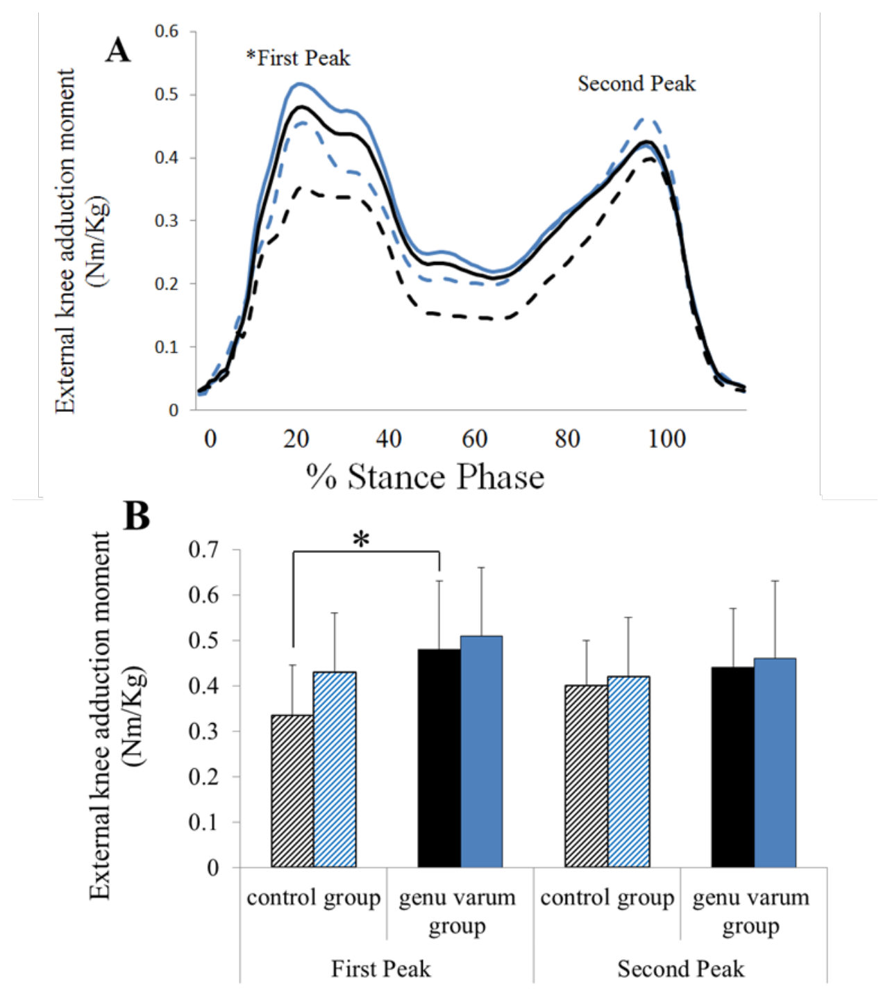
Fig. 1 illustrates the external knee adduction moment throughout the stance phase of walking in both groups under the two experimental conditions. No significant between-group differences were observed in the second peak of the external knee adduction moment under either condition (P > 0.05). However, before kinesiotaping, the genu varum group exhibited a significantly greater first peak of the external knee adduction moment than the control group (P = 0.029). Following kinesiotaping, the first peak increased in both groups, and the between-group difference was no longer statistically significant (Fig. 1B; P > 0.05).
Fig. 1. 1(A) Line graph of the knee external adductor moment throughout the entire stance phase for the control group pre-test: dashed black; control group in parentheses: solid black; and control group post-test: dashed blue; control group in parentheses: solid blue. (B) Bar chart of the quantified first and second peaks of the knee external moment for the control group pre-test: hashed black; control group in parentheses: solid black; and control group post-test: hashed blue; control group in parentheses: solid blue.
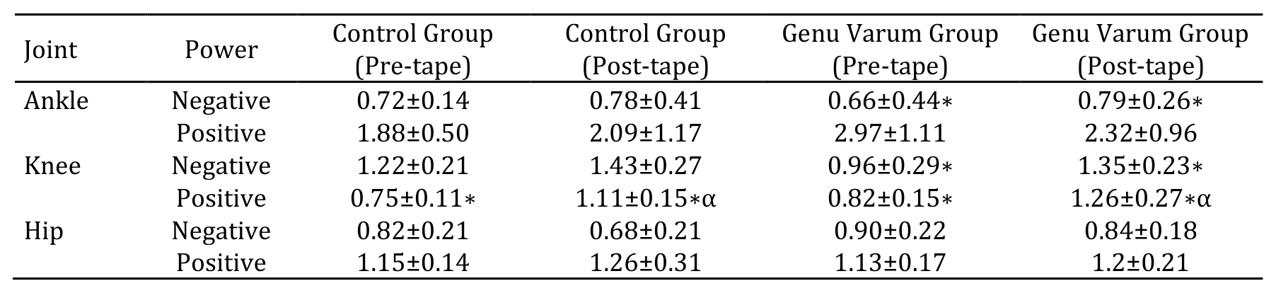
Table 1 presents the peak positive and negative joint power values at the hip, knee, and ankle during walking under both taping conditions. Following kinesiotaping, the genu varum group exhibited 27% greater positive knee joint power than the control group (P = 0.026). No significant between-group differences were observed for the remaining joint power variables (P > 0.05).
Within-group comparisons showed that kinesiotaping was associated with a 20% increase in the magnitude of negative ankle joint power in the genu varum group (P = 0.038). At the knee joint, positive power increased by 48% in the control group (P = 0.017). In the genu varum group, the magnitude of negative knee joint power increased by 40% (P = 0.025), while positive knee joint power increased by 53% (P = 0.012). No significant changes in hip joint power were observed following kinesiotaping (P > 0.05).
Table 1. 1Mean ± standard deviation of peak joint power (W/kg) at the hip, knee, and ankle during walking in different groups.
*Significant difference between taping conditions; αSignificant difference between the control group and the genu varum group.
Discussion
The present study investigated the immediate effects of kinesiotaping applied to the biceps femoris and vastus lateralis muscles on the external knee adduction moment and lower-limb joint power during walking in individuals with genu varum and healthy controls. Before kinesiotaping, individuals with genu varum exhibited a significantly greater first peak of the external knee adduction moment than healthy controls. This finding is consistent with previous studies reporting greater external knee adduction moments in individuals with genu varum (8, 9). The external knee adduction moment is commonly used as a biomechanical surrogate of medial knee compartment loading, although it does not directly quantify joint contact forces. Previous research has associated elevated knee adduction moments with knee pain and the development or progression of medial compartment osteoarthritis (10–12). However, the relationship between knee adduction moments and clinical outcomes is complex and may be influenced by individual anatomical and biomechanical characteristics.
Following kinesiotaping, the first peak of the external knee adduction moment increased numerically in both groups, and the previously significant between-group difference was no longer observed. However, the absence of a statistically significant between-group difference after taping does not necessarily indicate a significant treatment effect. Moreover, because the external knee adduction moment is associated with medial compartment loading, an increase in this variable cannot be interpreted as a beneficial mechanical adaptation without additional evidence. Therefore, the present findings do not support the initial hypothesis that kinesiotaping would reduce the external knee adduction moment in individuals with genu varum. Further studies incorporating appropriate group-by-condition interaction analyses are needed to clarify whether kinesiotaping produces different biomechanical responses in individuals with and without genu varum.
Regarding ankle joint power, kinesiotaping was associated with a significant increase in the magnitude of negative ankle power in the genu varum group. Negative joint power generally represents net mechanical energy absorption at a joint and is influenced by the interaction between joint moments and angular velocities. During the early stance phase of walking, eccentric activity of the ankle dorsiflexors contributes to controlling plantarflexion following initial contact (13). The observed increase in the magnitude of negative ankle power may therefore reflect altered mechanical energy absorption at the ankle. However, because the present study evaluated peak joint power rather than phase-specific energy absorption, the timing and functional significance of this change cannot be established. Furthermore, whether this alteration contributes to improved shock attenuation or a reduced risk of lower-limb injury remains uncertain.
Kinesiotaping was also associated with significant increases in the magnitude of negative knee joint power and positive knee joint power in individuals with genu varum. During walking, negative knee joint power is commonly associated with net mechanical energy absorption, particularly during weight acceptance, when eccentric quadriceps activity contributes to controlling knee flexion (14). Accordingly, the observed increase in the magnitude of negative knee power may indicate changes in the mechanical demands placed on the knee during gait. However, peak negative joint power alone does not establish improved shock absorption or reduced ground reaction forces. Similarly, the increase in positive knee joint power suggests altered net mechanical power generation but does not necessarily indicate improved walking efficiency or functional performance. Although kinesiotaping has been proposed to influence proprioceptive feedback and neuromuscular coordination (15), these mechanisms were not directly assessed in the present study. Therefore, the underlying mechanisms responsible for the observed changes in joint power remain unclear.
In contrast, kinesiotaping did not produce significant changes in hip joint power during walking. This finding suggests that the immediate biomechanical effects observed in the present study were more apparent at the knee and ankle than at the hip. During gait, hip joint power reflects the combined contributions of joint moments and angular velocities, with positive and negative power occurring during different phases of the gait cycle (16, 17). Because kinesiotaping was applied only to the biceps femoris and vastus lateralis muscles, the intervention may have had a more localized influence on lower-limb mechanics. Nevertheless, this interpretation remains speculative, as muscle activation patterns and interjoint coordination were not directly examined. The absence of statistically significant changes in hip joint power should also not be interpreted as evidence that kinesiotaping has no effect on hip mechanics.
Several limitations should be considered when interpreting these findings. First, the relatively small sample size and inclusion of only young male participants limit the generalizability of the results. Second, the absence of a sham-taping condition makes it difficult to distinguish the specific effects of kinesiotaping from nonspecific effects associated with tape application. Third, the potential influence of walking speed and the order of experimental conditions should be considered when interpreting changes in joint moments and power. In addition, the study focused on immediate biomechanical responses and did not assess long-term adaptations, clinical outcomes, or injury incidence. Future studies should incorporate sham-controlled designs, appropriate control of walking speed, and comprehensive analyses of joint kinetics and muscle activation. Longitudinal investigations are also needed to determine whether the observed biomechanical changes have meaningful clinical implications for individuals with genu varum.
Conclusion
This study demonstrated that individuals with genu varum exhibit higher external knee adduction torque (EKAT) and greater positive knee joint power during gait compared to healthy individuals. Since kinesio taping of the biceps femoris and vastus lateralis muscles modifies the external adductor moment of the knee, this may lead to a reduced risk of developing knee osteoarthritis in individuals with genu varum. Furthermore, because kinesiotaping of these muscles increases ankle and knee power, this tool may be considered an efficient and effective method for enhancing gait efficiency and improving walking mechanisms in individuals with genu varum. However, the use of kinesiotape did not have a significant effect on hip joint power during gait.
Ethical Considerations
Compliance with ethical guidelines
All participants provided written informed consent before participating in the study. The study was conducted in accordance with the ethical principles outlined in the Declaration of Helsinki. Participation was voluntary, and the confidentiality of participants' personal information was maintained throughout the study.
Funding
This research received no specific grant from any funding agency in the public, commercial, or not-for-profit sectors.
Authors' contributions
Morteza Madadi-Shad: Formal analysis, Supervision, Writing – original draft, Writing – review & editing.
Sara Bagheri: Writing – original draft, Writing – review & editing.
Mahdieh Irom: Investigation, Project administration.
Mohammadhossein Ghasemi: Conceptualization, Methodology, review & editing.
All authors read and approved the final version of the manuscript.
Conflicts of interest
The authors declare no conflict of interest.
Type of Study: Research |
Subject:
Special
Received: 2026/08/24 | Accepted: 2026/10/14 | Published: 2026/10/15
Received: 2026/08/24 | Accepted: 2026/10/14 | Published: 2026/10/15
References
1. Shibuya N, Kitterman RT, LaFontaine J, Jupiter DC. Demographic, physical, and radiographic factors associated with functional flatfoot deformity. The Journal of Foot and Ankle Surgery. 2014;53(2):168-172. [DOI:10.1053/j.jfas.2013.11.002]
2. Jafarnezhadgero A, Shad MM, Ferber R. The effect of foot orthoses on joint moment asymmetry in male children with flexible flat feet. Journal of Bodywork and Movement Therapies. 2018;22(1):83-89. [DOI:10.1016/j.jbmt.2017.04.007]
3. Madadi-Shad M, Farahpour N, Majlesi M. Immediate effects of anti-pronation foot orthoses with different inclination angles on ground reaction force components during walking. The Scientific Journal of Rehabilitation Medicine. 2019;8(4):92-102.
4. Farahpour N, Jafarnezhad A, Damavandi M, Bakhtiari A, Allard P. Gait ground reaction force characteristics of low back pain patients with pronated foot and able-bodied individuals with and without foot pronation. Journal of Biomechanics. 2016;49(9):1705-1710. [DOI:10.1016/j.jbiomech.2016.03.056]
5. Farahpour N, Jafarnezhadgero A, Allard P, Majlesi M. Muscle activity and kinetics of lower limbs during walking in pronated feet individuals with and without low back pain. Journal of Electromyography and Kinesiology. 2018;39:35-41. [DOI:10.1016/j.jelekin.2018.01.006]
6. Asghari V, Farahpour N, Robbins S, Moisan G, Majlesi M. Kinematic changes of lower-limb joints during walking in individuals with chronic ankle instability on flat and inclined surfaces. Journal of Sport Biomechanics. The Foot. 2026;12(2):302-319. [DOI:10.66224/JSportBiomech.12.2.302]
7. Jafarnezhadgero AA, Shad MM, Majlesi M. Effect of foot orthoses on the medial longitudinal arch in children with flexible flatfoot deformity: a three-dimensional moment analysis. 2017;55:75-80. [DOI:10.1016/j.gaitpost.2017.04.011]
8. Twomey D, McIntosh AJ. The effects of low arched feet on lower limb gait kinematics in children. The Foot. 2012;22(2):60-65. [DOI:10.1016/j.foot.2011.11.005]
9. Lin CJ, Lai KA, Kuan TS, Chou YL. Correlating factors and clinical significance of flexible flatfoot in preschool children. Journal of Pediatric Orthopaedics. 2001;21(3):378-382. [DOI:10.1097/01241398-200105000-00022]
10. Granacher U, Schellbach J, Klein K, Prieske O, Baeyens JP, Muehlbauer T. Effects of core strength training using stable versus unstable surfaces on physical fitness in adolescents: a randomized controlled trial. BMC Sports Science, Medicine and Rehabilitation. 2014;6(1):40. [DOI:10.1186/2052-1847-6-40]
11. Chen YC, Lou SZ, Huang CY, Su FC. Effects of foot orthoses on gait patterns of flat feet patients. Clinical Biomechanics. 2010;25(3):265-270. [DOI:10.1016/j.clinbiomech.2009.11.007]
12. Mohammad Zaheri R, Majlesi M, Fatahi A. Impact of lower-limb fatigue on kinetic risk factors for ACL injury during post-spike landings in volleyball athletes. Journal of Sport Biomechanics. 2026;11(4):466-484. [DOI:10.61882/JSportBiomech.11.4.466]
13. Tang SF, Chen CH, Wu CK, Hong WH, Chen KJ, Chen CK. The effects of total contact insole with forefoot medial posting on rearfoot movement and foot pressure distributions in patients with flexible flatfoot. Clinical Neurology and Neurosurgery. 2015;129:S8-S11. [DOI:10.1016/S0303-8467(15)30004-4]
14. Lin JL, Balbas J, Richardson EG. Results of non-surgical treatment of stage II posterior tibial tendon dysfunction: a 7- to 10-year follow-up. Foot & Ankle International. 2008;29(8):781-786. [DOI:10.3113/FAI.2008.0781]
15. Faul F, Erdfelder E, Lang AG, Buchner A. G*Power 3: a flexible statistical power analysis program for the social, behavioral, and biomedical sciences. Behavior Research Methods. 2007;39(2):175-191. [DOI:10.3758/BF03193146]
16. Nigg BM, Federolf P, Landry SC. Gender differences in lower extremity gait biomechanics during walking using an unstable shoe. Clinical Biomechanics. 2010;25(10):1047-1052. [DOI:10.1016/j.clinbiomech.2010.07.010]
17. Robertson DG, Caldwell GE, Hamill J, Kamen G, Whittlesey S. Research Methods in Biomechanics. 2nd ed. Champaign, IL: Human Kinetics; 2013. [DOI:10.5040/9781492595809]
18. Nigg BM, Mohr M, Nigg SR. Muscle tuning and preferred movement path: a paradigm shift. Current Issues in Sport Science. 2017;2:007. [DOI:10.15203/CISS_2017.007]
19. Boden BP, Torg JS, Knowles SB, Hewett TE. Video analysis of anterior cruciate ligament injury: abnormalities in hip and ankle kinematics. The American Journal of Sports Medicine. 2009;37(2):252-259. [DOI:10.1177/0363546508328107]
20. Yoshida M, Aoki N, Taniguchi K, Yoshida M, Katayose M. Kinematic analysis of the ankle joint on the side-hop test in subjects with ankle sprains. Translational Sports Medicine. 2018;1(6):265-272. [DOI:10.1002/tsm2.44]
21. Webster CA, Nussbaum MA. Localized ankle fatigue development and fatigue perception in adults with or without chronic ankle instability. Journal of Athletic Training. 2016;51(6):491-497. [DOI:10.4085/1062-6050-51.9.02]
22. Unnithan S, Thomas J. Not all ankle injuries are ankle sprains: case of an isolated cuboid stress fracture. Clinics and Practice. 2018;8(3):1093. [DOI:10.4081/cp.2018.1093]
23. Madadi-Shad M, Hanifeh MB, Momen HR. How far an arch support foot orthosis can affect lower limb joint angular accelerations during normal gait? Journal of Advanced Sport Technology. 2020;4(2):11-18.
24. Darvishi S, Majlesi M, Azadian E. Comparative analysis of center of pressure responses during defensive landing in professional and novice volleyball players. Journal of Sport Biomechanics. 2025;11(2):162-174. [DOI:10.61186/JSportBiomech.11.2.162]
25. Jafarnezhadgero A, Madadi-Shad M, Alavi-Mehr SM, Granacher U. The long-term use of foot orthoses affects walking kinematics and kinetics of children with flexible flat feet: a randomized controlled trial. PLOS ONE. 2018;13(10):e0205187. [DOI:10.1371/journal.pone.0205187]
26. Nester CJ, Van Der Linden ML, Bowker P. Effect of foot orthoses on the kinematics and kinetics of normal walking gait. Gait & Posture. 2003;17(2):180-187. [DOI:10.1016/S0966-6362(02)00065-6]
| Rights and permissions | |
 |
This work is licensed under a Creative Commons Attribution-NonCommercial 4.0 International License. |

Journal of Sport Biomechanics
Department of Sport Biomechanics, Faculty of Humanities, Islamic Azad University, Hamedan Branch, Prof. Mousivand Blvd, Imam Khomeini Blvd, Hamedan, Iran.
Journal Tel: +98 81 34494042
Website: http://biomechanics.iauh.ac.ir
Email: sportbiomechanics@iauh.ac.ir
